

How Are Dna Fragments Separated Using Gel Electrophoresis
Scientists use gel electrophoresis to separate molecules based on their size and electric charge. Gel electrophoresis tin separate fragments of DNA that were cut with restriction enzymes, creating a visual map of fragment size that's piece of cake to translate. Or scientists may apply gel electrophoresis to separate a protein they want to report from other proteins in a cell.
1 of the advantages of gel electrophoresis is that scientists can separate several samples side by side and then they can compare them. The comparison of separated DNA molecules is the basic method behind the Deoxyribonucleic acid fingerprints that forensic scientists employ to compare samples from offense scenes with those of suspects.
Scientists conduct gel electrophoresis by inserting molecules such as Deoxyribonucleic acid into little pockets called wells within a slab of gel. They then place the gel in a box called an electrophoresis sleeping accommodation that'south filled with a salty, electricity-conducting buffer solution.
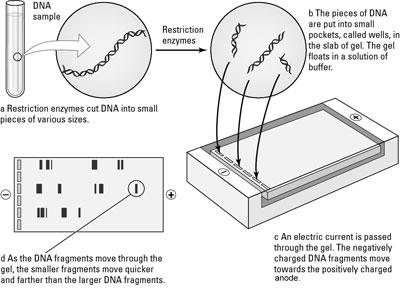
The DNA molecules, which have a negative charge, move toward the gel box'southward positive electrode because opposite charges concenter. When the scientists run an electrical current through the gel, the gel becomes like a racetrack for the DNA molecules equally they try to get to the positively charged end of the box.
When the power is turned off, all the DNA molecules terminate where they are in the gel, and the scientists stain them. The stain sticks to the DNA, creating stripes called bands. Each ring represents a collection of Dna molecules that are the same size and stopped in the same identify in the gel.
In social club to work with the information from the gel more than easily, scientists can make an identical copy of the gel by transferring the Dna molecules to a thin sheet of nylon or nitrocellulose, a strong but flexible material that binds to DNA. This procedure is called making a blot of the gel. A blot on a thin, flexible material can exist handled, whereas the original slab of gel can crevice and intermission.
-
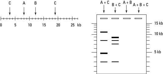
The post-obit effigy shows a linear piece of viral Dna that's 27 kb long and has restriction sites for the enzymes A, B, and C. After the viral DNA was cut with various combinations of restriction enzymes, the resulting Deoxyribonucleic acid fragments were separated using gel electrophoresis.
Based on the information given in the figure, what pattern of bands would you predict in the lane of the gel marked A + B, which would be loaded with many copies of viral Dna cut with both enzymes A and B?
-
If yous treated the viral DNA with all three restriction enzymes — A, B, and C — and then separated the fragments using gel electrophoresis, what blueprint of bands would announced in the last lane of the figure?
-
Bands would announced at the positions indicating three, five, 8, and xi kb
-
Bands would appear at the positions indicating four, 8, and fifteen kb
-
Bands would appear at the positions indicating 3, 7, viii, and 9 kb
-
Bands would appear at the positions indicating 3, 4, 5, 7, and 8 kb
-
-
The open up rectangles at the superlative of the gel in the figure represent the wells. Towards which end of the gel would the positive electrode be located?
-
The top
-
The lesser
-
-
The open up rectangles at the top of the gel in the figure represent the wells. If yous were to run a sample of uncut viral DNA through this gel for the same amount of time that the other samples were run, at which cease of the gel would yous expect to observe your band?
-
The top, near the wells
-
The bottom, away from the wells
-
The post-obit are the answers to the practice questions.
-
The viral Dna has a restriction site for enzyme A at a position 8 kb from the left end of the Dna. So, cut with enzyme A will generate some fragments that are 8 kb in length.
The viral Deoxyribonucleic acid likewise has a restriction site for enzyme B at a position just iv kb from the restriction site for enzyme A, so cut with restriction enzyme B will produce some fragments that are 4 kb in length. The cut with enzyme B will also produce remainder fragments that stretch from the restriction site for B all the way to the correct finish of the viral Deoxyribonucleic acid. These residue fragments will be fifteen kb in length.
To double-check your work, y'all can add up the lengths of your predicted fragments: 8 + 4 + 15 = 27, which is the correct length of the entire slice of viral Dna. To make up one's mind the position of the bands on the gel, look at the size labels along the right side of the gel. You would predict a ring of DNA fragments to appear level with the 4 kb marker, the 8 kb marker, and the 15 kb marker. All these bands should comprise the same numbers of fragments and then they should appear in equal thickness on the gel.
-
The reply is 4. Bands would appear at the positions indicating 3, four, 5, 7, and eight kb.
If you cut the viral DNA with all three enzymes, you'll make four cuts, resulting in five fragments.
-
The answer is ii. The lesser.
DNA, which is negatively charged, is loaded into the wells. The positive electrode is located at the far end, abroad from the Dna, so that it volition attract the Deoxyribonucleic acid from a distance, pulling it through the gel.
-
The answer is 1. The top, about the wells.
Uncut pieces of viral DNA would exist longer than any of the fragments fabricated by brake enzymes (they'd exist the entire 27 kb long), so they would travel a shorter distance than any of the fragments.
Nigh This Commodity
This article tin can exist found in the category:
- Biological science ,

How Are Dna Fragments Separated Using Gel Electrophoresis,
Source: https://www.dummies.com/article/academics-the-arts/science/biology/how-biologists-separate-molecules-with-gel-electrophoresis-168989/
Posted by: davisonated.blogspot.com
0 Response to "How Are Dna Fragments Separated Using Gel Electrophoresis"
Post a Comment